Malaria-parasites resistant to treatment and detection have emerged in Ethiopia

Scientists have detected new strains of Plasmodium falciparum parasite in Ethiopia that are both resistant to current treatments and escape detection by common diagnostic tests — a development that could increase cases and deaths from malaria and make eliminating the persistent disease an even greater challenge[1].
Earlier, scientists had found in Uganda, Tanzania and Rwanda strains of the parasite that causes malaria that were resistant to most available antimalarial drugs; and separately, malaria parasites resistant to diagnostic tests had emerged in the Horn of Africa.

Those parasites have been spreading independently of one another, but the new study is the first published report to confirm the prevalence of this type of double-resistant malaria strain, said study author Jeffrey Bailey, an associate professor of translational research and pathology and laboratory medicine at Brown University.

“Now we're essentially seeing the worst-case scenario: parasites with the mutation that make them resistant to treatment have also picked up the chromosomal deletions that make them invisible to the diagnostic tests,” Bailey said. “This means that it will be harder to detect people who are infected, and then when infected people are treated with antimalarial drugs, that may not work to stop them from spreading the disease.”

The first-line malaria treatment recommended by the World Health Organization is a combination therapy involving artemisinin-based drug compounds, which tend to be very effective in preventing death and reducing transmission. The mutations now detected in Africa provide resistance to artemisinin.

They found that 8.2% of drug-resistant parasites also carried the deletions of the protein-expressing gene (the candidate artemisinin partial-resistance kelch13 622I mutation) that made them undetectable by the diagnostic tests[2].

While in Ethiopia the overall incidence of malaria is low, the disease remains endemic in 75% of the country, with 65% of the population at risk. More than 5 million episodes of malaria occur each year.

“The spread of these parasites will certainly make eliminating malaria in Ethiopia and elsewhere in Africa more difficult and will likely lead to increased cases and deaths,” Bailey said.

[1] Fola et al: Plasmodium falciparum resistant to artemisinin and diagnostics have emerged in Ethiopia in Nature Biology – 2023. See here.
[2] Fola et al: Clonal spread of Plasmodium falciparum candidate artemisinin partial resistance Kelch13 622I mutation and co-occurrence with pfhrp2/3 deletions in Ethiopia in MedRχiv - 2023

Malaria and toxic bacteria

Mosquitoes might be infected with all sorts of pathogens, such as viruses and bacteria. When you get stung at night, the mosquito will inject a tiny amount of their saliva, which contains a specialized, potent cocktail of molecules that numb the pain of the bite and stop the blood from clotting. But, together with that saliva, the pathogens also enter your bloodstream.
Still, some bacteria seem to have a detrimental effect on the mosquito itself.

Scientists noticed that some laboratory colonies of mosquitoes were incapable of transmitting malaria parasites[1]. These insects also harboured a few cells of a bacterium called Delftia tsuruhatensis TC1, which produces a toxic alkaloid called harmane. Bacteria-produced harmane inhibited the development of female Plasmodium falciparum parasite gametes in the mosquito gut.

Harmane was found to be a contact poison that could also cross the mosquito cuticle to kill developing malaria parasites. The data suggests that the bacteria can reduce a mosquito's parasite load by up to 73%.

Contained field trials in Burkina Faso, coupled with modeling studies, showed that the bacterium has the potential to be deployed in mosquito breeding sites as a component of malaria control.

Harmane is a heterocyclic amine found in a variety of foods, including coffee[2]. Harmane is also present in tobacco smoke[3].

[1] Huang et al: Delftia tsuruhatensis TC1 symbiont suppresses malaria transmission by anopheline mosquitoes in Science – 2023
[2] Herraiz, Chaparro: Human monoamine oxidase enzyme inhibition by coffee and β-carbolines norharman and harman isolated from coffee in Life Sciences - 2006
[3] Herraiz, Chaparro: Human monoamine oxidase is inhibited by tobacco smoke: beta-carboline alkaloids act as potent and reversible inhibitors in Biochemical and Biophysical Research Communications - 2005

Malaria species jumps from apes to humans

Malaria is a disease caused by the protozoa of plasmodium species that is transmitted via the infected female Anopheles mosquito. Traditionally, there are four plasmodium species that cause natural human malaria infection: Plasmodium falciparum, Plasmodium vivax, Plasmodium ovale, and Plasmodium malariae.
[Image: Rossche - Long-tailed macaque]

Just four? Not anymore.

Several simian plasmodium species have been reported to be able to infect humans. At least seven species of monkey malaria have been reported as transmissible to man by mosquito bite: Plasmodium cynomolgi, Plasmodium brasilianum, Plasmodium eylesi, Plasmodium knowlesi, Plasmodium inui, Plasmodium schwetzi, and Plasmodium simium. The first known incident occured in 1960, when two laboratory workers became accidentally infected with Plasmodium cynomolgi via an Anopheles mosquito[1].

Plasmodium cynomolgi predominately causes malaria in macaque monkeys but is – as mentioned above - also known to cause experimental and rare natural zoonotic infections in humans. Recently, however, Plasmodium cynomolgi has made the zoonotic jump and started naturally infecting humans[2].

The first naturally acquired human infection with Plasmodium cynomolgi occurred at the start of 2011 in a 39-year-old Malay woman from the east coast of Peninsular Malaysia[3]. Behind her house there is a small, forested area with occasional sightings of long-tailed macaques (Macaca fascicularis).

Since then, there have been another symptomatic infection of a tourist travelling to Malaysia, cases in Malaysian Borneo, two additional Malaysian cases and multiple asymptomatic infections in Cambodia.

The significance of Plasmodium cynomolgi that is transmissible cannot be ignored nor underestimated.

[1] Eyles et al: Vivax-type malaria parasite of macaques transmissible to man in Science – 1960
[2] Bykersma: The New Zoonotic Malaria: Plasmodium cynomolgi in Tropical Medicine and Infectious Diseases – 2021. See here.
[3] Ta et al: First case of a naturally acquired human infection with Plasmodium cynomolgi in Malaria Journal – 2014. See here.

Link found between malaria parasites, humans, and apes

Scientists have solved a century-old mystery about the evolutionary links between malaria parasites that now infect humans and chimpanzees.
They have discovered that the parasite Plasmodium malariae — one of six species that spreads malaria among humans — originated in African apes before evolving to infect people.

While it is often associated with mild disease, if left untreated Plasmodium malariae can cause long-lasting, chronic infections that may last a lifetime, researchers say.

The evolutionary puzzle has its origins in the 1920s when scientists identified chimpanzees infected by parasites that appeared identical to Plasmodium malariae under a microscope[1].

It was thought both parasites belonged to the same species, but – until now — this could not be verified as the genetic make-up of the chimpanzee strain had never been studied. Recently, scientists have used novel techniques to study the parasites' mitochondrial DNA (mtDNA)[2].

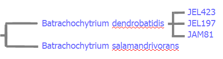
They have found that there are, in fact, three distinct species. One species, Plasmodium malariae, infects mainly humans, while the two others infect apes.

One of the two ape-infecting parasites was found in chimpanzees, gorillas and bonobos across Central and West Africa. This previously unknown species, provisionally called M2, is only distantly related to the human parasite.

The second ape parasite, provisionally called M1-like, is much more closely related to the lineage that infects humans, but exhibits little evidence of genetic exchange with it, and so likely represents a separate species.
This enabled researchers to make detailed comparisons of the genetic diversity of the two species. This revealed that the human malaria parasite population went through some sort of genetic bottleneck, where its population temporarily shrank and most of its genetic variation was lost. A likely explanation for this is that Plasmodium malariae was originally an ape parasite, but a small number of parasites switched hosts to begin infecting humans, the team speculates.

Lead author Dr. Lindsey Plenderleith said: "Among the six parasites that cause malaria in humans, Plasmodium malariae is one of the least well understood. Our findings could provide vital clues on how it became able to infect people, as well as helping scientists gauge if further jumps of ape parasites into humans are likely."

[1] Reichenow: Über das Vorkommen der Malariaparasiten des Menschen bei den Afrikanischen Menschenaffen in Centralblatt für Bakteriologie und Parasitenkunde - 1920
[2] Plenderleith et al: Zoonotic origin of the human malaria parasite Plasmodium malariae from African apes in Nature Communications. 2022. See here.

Avian Malaria

All variants of malaria is caused by infection with parasites in the genus Plasmodium and there are quite a number of these pesky parasites. The genus Plasmodium consists of over 200 species, generally described on the basis of their appearance in blood smears of infected vertebrates. These species have been categorized on the basis of their morphology and host range into 14 subgenera.
In humans, malaria is caused by six Plasmodium species: Plasmodium falciparum (~75%), Plasmodium vivax (~20%), Plasmodium malariae, Plasmodium ovale curtisi, Plasmodium ovale wallikeri, and Plasmodium knowlesi.

But some species of Plasmodium also infect birds and then causes avian malaria. Avian malaria is usually caused by Plasmodium relictum. Yes, there are several other species of Plasmodium that infect birds, such as Plasmodium anasum and Plasmodium gallinaceum, but these are of lesser importance

Avian malaria is found worldwide, with some important exceptions. Usually, it does not kill the infected birds. However, in areas where avian malaria is recently introduced, such as the islands of Hawaiʻi, it can be devastating to birds that have lost evolutionary resistance over time[1].

The parasite seems to be evolving to evade immunity, while it also seems to be able to expand its geographical range[2].

[1] McClure et al: The role of native and introduced birds in transmission of avian malaria in Hawaii in Ecology – 2020
[2] Theodosopoulos et al: A highly invasive malaria parasite has expanded its range to non-migratory birds in North America in Biology Letters – 2021

WHO recommends malaria vaccine for children

The World Health Organization (WHO) is recommending widespread use of the RTS,S/AS01 (RTS,S) malaria vaccine (trade name Mosquirix) among children in sub-Saharan Africa and in other regions with moderate to high Plasmodium falciparum malaria transmission. The recommendation is based on results from an ongoing pilot programme in Ghana, Kenya and Malawi that has reached more than 800 000 children since 2019.
“This is a historic moment. The long-awaited malaria vaccine for children is a breakthrough for science, child health and malaria control,” boasted WHO Director-General Dr Tedros Adhanom Ghebreyesus. “Using this vaccine on top of existing tools to prevent malaria could save tens of thousands of young lives each year.”

Malaria remains a primary cause of childhood illness and death in sub-Saharan Africa. More than 260 000 African children under the age of five die from malaria annually.

"For centuries, malaria has stalked sub-Saharan Africa, causing immense personal suffering,” said Dr Matshidiso Moeti, WHO Regional Director for Africa. “We have long hoped for an effective malaria vaccine and now for the first time ever, we have such a vaccine recommended for widespread use. Today’s recommendation offers a glimmer of hope for the continent which shoulders the heaviest burden of the disease and we expect many more African children to be protected from malaria and grow into healthy adults.”

WHO recommends that in the context of comprehensive malaria control the RTS,S/AS01 malaria vaccine be used for the prevention of Plasmodium falciparum malaria in children living in regions with moderate to high transmission as defined by WHO. RTS,S/AS01 malaria vaccine should be provided in a schedule of 4 doses in children from 5 months of age for the reduction of malaria disease and burden.
Next steps for the WHO-recommended malaria vaccine will include funding decisions from the global health community for broader rollout, and country decision-making on whether to adopt the vaccine as part of national malaria control strategies.

Source.

[Update 02 October 2023] Oxford R21/Matrix-M™ malaria vaccine receives WHO recommendation paving the way for global roll-out. See here.

Researchers develop vaccine for babesiosis

Griffith University (Australia) researchers have developed a new vaccine to treat human babesiosis, a deer tick-transmitted parasitic disease closely related to malaria[1]. An infection with Babesia microti, a parasitic blood-borne piroplasm, casues babesiosis.
Lead researcher Professor Michael Good AO, said the research team including PhD candidate Hanan Al-Nazal and researcher Dr Danielle Stanisic, developed a whole parasite Babesia vaccine that acts as a universal vaccine, inducing immunity against different human Babesia species. The vaccine is in pre-clinical trails.

“Babesiosis affects the red blood cells similar to malaria in humans and animals and presents as a flu-like illness and anaemia. People most at risk of severe disease are the elderly, the immunosuppressed and those without spleens. It also affects those who need blood transfusions,’’ Professor Good said.

“In pre-clinical studies we have shown this vaccine can kill the parasite and induce a protective immune response. The immune response is tied to two crucial aspects of the immune system – T Cells and macrophages (which clear bacteria and other germs).”

The vaccine is delivered using a liposomal platform (where the killed parasite is contained in a lipid vesicle). The advantage of liposomes is that they can be freeze-dried so they are suitable for transporting into the field.

Dr Stanisic said as far as they were aware, this was the first time a whole parasite vaccine for babesia had been developed.

[1] Al-Nazal et al: Pre-clinical evaluation of a whole-parasite vaccine to control human babesiosis in Cell Host and Microbe - 2021. See here.

Amphibian collapses increase malaria outbreaks

The Amphibian Chytrid Fungus (Batrachochytrium dendrobatidis) is a highly contagious fungus that infects the skin of amphibians, blocking them from breathing and is eventually fatal. Ponds hit by the fungus are quickly choked by dead or almost dead frogs. It is so deadly and so easily spread that it has already caused the extinction of an estimated 90 species of frogs, and reduced the populations of another 124 species by over 90%. This makes it “the most destructive pathogen ever described by science,” as Wendy Palen, biologist, wrote in an article last year. One study traced its origin to Korea in the 1950s, after which humans spread it across the world.
[Image: Geoff Gallice]

Frogs and other amphibians consume a large amount of mosquitoes. If you remove a huge number of these amphibians from their habitat, you can image what must happen to malaria rates.

Researchers cross-referenced dates of Amphibian Chytrid Fungus-driven amphibian decline in different parts of Costa Rica and Panama with changes in malaria incidence in those same places[1]. The Amphibian Chytrid Fungus swept across these two countries over the course of about twenty-five years, starting in north-western Costa Rica and then slowly, but surely progressing south and east. They found that, generally, around a year after the Amphibian Chytrid Fungus entered a county, malaria cases began to increase. They continue to rise for two years, then stay at that higher level for six more, before beginning to attenuate nine years after the fungus arrived.

“For the six years our estimated effect of amphibian decline is at its highest, the annual expected increase in malaria ranges from 0.76-1.0 additional cases per 1,000 population,” they write, which makes up “a substantial share of cases overall.” This rate of increase in Costa Rica’s capital, San Jose, would translate to 1000 more cases there.

While it’s possible that something besides a causal relationship could explain these numbers, it is “extremely unlikely,” the authors write.

“The results in our paper suggest that some policies, such as amphibian conservation policies or the regulation of wildlife trade, could have benefits for human health which are not currently accounted for,” fellow-author Joakim Weill, says.

Whatever happens, Mother Nature has already found another way of killing amphibians, because a recently described second species, Batrachochytrium salamandrivorans, also cause chytridiomycosis and death in salamanders and newts in The Netherlands[2].

[1] Springborn et al: Amphibian collapses exacerbated malaria outbreaks in Central America in MedrΧiv - 2020
[2] Martel et al: Batrachochytrium salamandrivorans sp. nov. causes lethal chytridiomycosis in amphibians in PNAS - 2013

Artemisinin resistant Malaria strain reported in Africa

Almost 3.2 billion people (virtually half the world’s population) in 89 countries are at risk of contracting Malaria. Every year, over 200 million cases and over 400,000 deaths are recorded.

For more than 15 years, treatment of malaria episodes (typical cycles of the disease alternating between fever, shivering and chills, and severe sweating) caused by Plasmodium falciparum has depended on artemisinin-based combination therapies (ACTs), which combine a fast-acting artemisinin derivative and a partner drug with a long half-life.
[Artemisia annua]
Resistance to artemisinin, the main component of the current antimalarial treatments recommended by WHO, is already widespread in South-East Asia, but it had not previously been described in Africa. A major concern is that these resistant parasites will spread through Sub-Saharan Africa, the continent most affected by malaria (>90% of all cases).

Since 2008, parasites capable of resisting artemisinin derivatives in South-East Asia have become increasingly prevalent. This resistance, which leads to a delay in the clearance of parasites from the bloodstream of individuals treated with an ACT, is currently a serious threat that may hinder efforts to tackle the disease.

Since 2014, the distribution of artemisinin resistance has been monitored based on the detection of mutations in parasites. Currently, the most widespread resistant parasites in South-East Asia have the C580Y mutation[1]. Recently, C580Y mutant parasites have also been detected in Guyana and Papua New Guinea.
[Plasmodium falciparum]
Scientists recently identified the first signs of emergence of artemisinin-resistant mutant parasites in Africa. The results describe significant proportions of parasites carrying the R561H mutation in two locations 100 kilometers apart[2]. Whole-genome sequencing of these parasites indicates that the R561H mutants were, unexpectedly, not acquired from Asian parasites.

The fact, that this resistant strain has spread between several places in Rwanda and its ability to resist artemisinin in vitro, has major public health implications. There is a risk that over time they will acquire the ability to resist the partner drugs used in ACTs. This would mean that the only available treatments would become ineffective, as has occurred in South-East Asia.

[1] Zaw et al: Importance of kelch 13 C580Y mutation in the studies of artemisinin resistance in Plasmodium falciparum in Greater Mekong Subregion in Journal of Microbiology, Immunology and Infection – 2019. See here.
[2] Uwimana et al: Emergence and clonal expansion of in vitro artemisinin-resistant Plasmodium falciparum kelch13 R561H mutant parasites in Rwanda in Nature Medicine – 2020. See here.

Malaria and Coffeewood

Malaria parasites have the annoying habit of circumventing our medicine to combat malaria. They mutate, become resistant and medication like quinine or artimisine are becoming more and more ineffective.
The current main therapy is ACT (artemisinin-based combination treatment), introduced when the parasite became resistant to chloroquine, a quinine derivative. Now artemisinin resistance is becoming increasingly common and no new class of antimalarial has been introduced since 1996.

That means that the search is on for other therapies. One of these is coffeewood (Caesalpinia pluviosa)[1].

Coffeewood is a legume within the Fabaceae family with numerous local medicinal uses, many of which have some rational basis. The plant is antiviral, antimicrobial, anti-inflammatory and antioxidant. Apparently, it is also anti-malarial. In previous research, the crude extract proved inactive. The current research started in vitro testing various extracts against Plasmodium. Finding the hoped for activity, research moved to in vivo research in infected mice.

In the in vitro test, two fractions were significantly effective. The crude extract was not. In mice, the crude extract was somewhat effective, though not as effective against chloroquine resistant malaria. The ethanol extract was effective against both. What’s more, it was synergistic with the artemisinin based drug artesunate, so the two together are more effective than the combined effect of both. The plant extract alone was around 50% effective, artesunate around 60% and the combination around 80%.

Chemical analysis showed that a novel active molecule, most likely an isomer of quercetin, appears to be the most active compound against malaria.

[1] Kayano et al: In vitro and in vivo assessment of the anti-malarial activity of Caesalpinia pluviosa in Malaria Journal - 2011. See here.

Coronavirus and Chloroquine?

Recently, one non-peer-reviewed publication claimed that a combination of hydroxychloroquine and azithromycin might be effective for treating the novel Coronavirus or Covid-19[1].
Hydroxychloroquine is a derivative of chloroquine, a common antimalarial drug. It is also used to treat amoebic liver abscesses when other drugs used for such infections are not working. These drugs also mildly suppress the immune system, which is why they are used as part of the treatment of some autoimmune disorders, such as lupus erythematosis or rheumatoid arthritis. It’s been known that chloroquine has some antiviral activity against certain viruses.

One thing that should be understood is that these are not entirely benign drugs. They have a number of side effects and adverse reactions. In addition to more mild side effects, such as nausea, headache, loss of appetite, and diarrhea, there are two more severe potential side effects. One is that long term use of these drugs can damage the retina and lead to macular degeneration, which is why patients taking these drugs long term need regular ophthalmological examinations. They can also affect the heart.

Another concern was that it is easy to overdose on hydroxychloroqine, its therapeutic window (the difference between the lowest effective dose and doses that will cause toxicity, in this case cardiac toxicity) being narrow. 

The other drug in the combination, azithromycin is a common antibiotic, used to treat a number of infections. It can also be used to treat malaria. It has few adverse side effects, but it shares one with hydroxychloroquine: it affects the heart. The FDA issued a warning in 2013 that azithromycine “can cause abnormal changes in the electrical activity of the heart that may lead to a potentially fatal irregular heart rhythm.”

The study by Gautret et al. regarding the effect of hydroxychloroquine and azithromycin on coronavirus is not a randomized study and had only 36 patients. There is no evidence from this paper that these two drugs made any difference in the clinical outcomes of these patients.

This paper almost certainly would have failed peer review, given that it is not randomized, doesn’t do a proper intention-to-treat analysis, and has a whole lot of missing datapoints among the control subjects.

So, could hydroxychloroquine and chloroquine be effective drugs to treat and/or prevent coronavirus? It is a possiblity. There exists a plausible mechanism by which the drugs could inhibit viral replication, plus in vitro evidence of antiviral activity.

[Update 08 April 2020] Swedish hospitals abandon trial of promising malaria drug chloroquine for coronavirus patients after it caused them blinding headaches, vision loss and agonising cramps. See here.

[1] Gautret et al: Hydroxychloroquine and azithromycin as a treatment of COVID-19: results of an openlabel non-randomized clinical trial in [not officially published] - 2020. See here (pdf).

Coffee as Treatment for Malaria?

During the American Civil War (1861-1865), between the North (the Union) and the South (the Confederacy), unreliable supply and high cost of quinine forced the Confederate Army to use alternative treatments for malaria.
Many quinine substitutes were mentioned in the literature of the time, but relatively few were advocated by Confederate officials and even fewer are described in surviving records. Medical supply officers often issued substitute remedies when quinine was requisitioned. Most alternative treatments were made from indigenous plants such as dogwood (Cornus florida), willow (Salix spp.) and tulip tree (Liriodendron tulipifera). High hopes were held for Georgia bark (Pinckneya pubens), which was thought to be closely related to cinchona. However, documentation of the effectiveness of these quinine substitutes remains scant.

The earliest commonly used quinine substitutes were made from dogwood bark, alone or in combination with other barks. A batch of the most well-known recipe, compound tincture of indigenous barks, was to be substituted as far as practicable for quinine. It was made by soaking 2 pounds of mixed dried bark (30% dogwood, 30% tulip tree, and 40% willow) in one gallon of whiskey for two weeks.

There had been rare reports in the literature about treating malaria with unroasted coffee[1]. One physician, for example, wrote in 1821 about his success using a strong decoction of “unburnt” coffee to treat a child with intermittent fever[2] and an article in Scientific American claimed that “a singular peculiarity of coffee” was that “if used in its raw state ... it produces febrifuge effects[3].”

Coffee, however, was scarce in the Confederacy, especially late in the war, and its use as a quinine substitute might have been impractical. Because roasted coffee was popular in both the North and South, its value (or worthlessness) as an antimalarial agent should have been apparent well before 1865.

So, the question remains whether coffee, unroasted or not could be a viable alternative for quinine. The answer is probably not, because no scientific research has ever been published regarding the anti-malarial potential of coffee.
But recent research has revealed that Aspidosperma ramiflorum, a tree growing in the Brazilian rainforests, does have cytotoxic effects towards malaria parasites[4]. One of its local names is peroba-café. The name derives from the language of indigenous Tupi, where iperoba means 'bitter bark'. Which means we have 'bitter bark coffee'.

Reality often plays games with history.

[1] Wood, Bache: The Dispensatory of the United States of America, Editon 11 - 1858
[2] Baxter: Case of intermittent fever, treated with coffee. Medical Repository – 1821
[3] Coffee. Scientific American, Issue 3 – 1848
[4] Aguiar et al: Aspidosperma (Apocynaceae) plant cytotoxicity and activity towards malaria parasites. Part II: experimental studies with Aspidosperma ramiflorum in vivo and in vitro in Memórias do Instituto Oswaldo Cruz – 2015. See here.

Failed psoriasis medicine a potential malarial cure

A failed study into a remedy for skin disease psoriasis may be the breakthrough for a cure for malaria, according to RadboudUMC in Nijmegen. One of the Dutch researchers found a reference to possibly tackling malaria in the same way the failed psoriasis remedy worked in a book from 1946, and the method shows promise, the Dutch newspaper Telegraaf reported.
"The potential medicine, a pantothenamide molecule, is very similar to a molecule that occurs naturally in the malaria parasite. As a result, the parasite uses the medicine in almost the same way in the metabolism. With the big difference that this molecule causes problems for the metabolism of the single-cell malaria parasite, which dies at a result", RadboudUMC said.

Even a single dose of the medicine already seems to have an effect on the malaria parasite, according to one of the researchers[1]. "Moreover, it is cheap to produce and stops the transfer of the malaria parasite from humans to mosquitos. With that, if it grows into a fully-fledged medicine, it can also contribute to the eradication of malaria."

Of course, the medicine is still a way away from the market. Clinical research must first be done to determine its safety and effective doses.

[1] Schalkwijk et al: Antimalarial pantothenamide metabolites target acetyl–coenzyme A biosynthesis in Plasmodium falciparum in Science Translational Medicine - 2019

Multi-drug resistant malaria strain spreads in Southeast Asia

Genomic surveillance has revealed that malaria resistance to two first-line antimalarial drugs has spread rapidly from Cambodia to neighbouring countries in Southeast Asia. Researchers discovered that descendants of one multi-drug resistant malaria strain are replacing the local parasite populations in Vietnam, Laos and northeastern Thailand[1]. They also found the resistant strain has picked up additional new genetic changes, which may be enhancing resistance even further.

Over the last decade, the first-line treatment for malaria in many areas of Asia has been a combination of two antimalarial drugs, known as DHA-PPQ (Dihydroartemisinin and Piperaquine). However, a previous study identified a strain of malaria that had become resistant to this treatment. Researchers found that this resistant strain, named KEL1/PLA1, because of its combination of genetic mutations that cause resistance, had spread across Cambodia under the radar between 2007 and 2013.

In their study of malaria parasites in Southeast Asia, the team sequenced and analysed the DNA of 1,673 Plasmodium falciparum parasites, taken from the blood of malaria patients between 2008 and 2018. Their analysis, focusing on the KEL1 and PLA1 gene variants, revealed that the situation had got much worse since 2013. The multidrug resistant KEL1/PLA1 parasites had spread internationally, replacing local malaria parasites, in some regions making up more than 80 per cent of the parasites analysed.

The spread is likely to have occurred because resistant parasites had an evolutionary advantage, as DHA-PPQ was the first-line treatment in most of these areas. This killed other malaria strains but was less effective against KEL1/PLA1 malaria.

The researchers discovered that not only had this resistant strain spread geographically, but it had evolved and picked up new mutations in the chloroquine resistance transporter gene (crt). A related paper on clinical outcomes revealed that these crt mutations were associated with complete treatment failure of DHA-PPQ[2]. This supported the finding that the resistance had not only spread, but worsened as the parasite evolved under further drug pressure.

[1] Hamilton et al: Evolution and expansion of multidrug-resistant malaria in southeast Asia: a genomic epidemiology study in The Lancet – 2019
[2] Ménard, Fidock: Accelerated evolution and spread of multidrug-resistant Plasmodium falciparum takes down the latest first-line antimalarial drug in southeast Asia in The Lancet – 2019

Fever-Free Soft Drinks against Malaria

Tonic water manufacturer Fever-Tree is hoping to accelerate the fight against malaria with a new social media campaign, called Raise Your Glass, Erase Malaria.
From 22 April to 31 May 2019, Fever-Tree is calling on imbibers around the world to raise a glass to malaria’s demise and share an image of the moment on social media, tagging @FeverTreeMixers and #MalariaMustDie.

For every photo shared across Instagram, Twitter and Facebook, Fever-Tree will donate £5 (US$6) to UK charity Malaria No More.

The initiative is part of the global campaign Malaria Must Die, So Millions Can Live, which was developed by Malaria No More in the run up to World Malaria Day (25 April).

The social media campaign is the latest in Fever-Tree’s continued fight against malaria, which has so far seen the mixer manufacturer partner with Malaria No More to help convene a Malaria Summit in London. Following the summit, 53 Commonwealth nations made a commitment to halve malaria in the Commonwealth over the next five years.

Earlier this year, Fever-Tree committed to a £1 million partnership with Malaria No More, which will fund a number of initiatives aimed at raising awareness and support for the charity.

Quinine and Leg Cramps

Muscle cramp or leg cramp is a recurrent and painful condition and a common complaint among the elderly. It is commonly nocturnal and can disturb a good night's sleep. No specific cause can be identified and therapy is mostly symptomatic.
Quinine sulfate (200-300 mg at bedtime) is widely used as an effective therapy for idiopathic nocturnal leg cramps[1]. Several studies have questioned the effectiveness of quinine in leg cramps, whereas other studies have shown significantly more benefit with use of quinine in reducing the frequency and severity of cramps compared with placebo or vitamin E.

However, the mechanism of this effect is obscure. Quinine appears to decrease the excitability of the motor end plate, thereby reducing the muscle contractility.

New research has found that long term use of quinine, prescribed off label for leg cramps, muscle cramps or restless leg syndrome, is associated with increased mortality[2]. Therefore, it is not recommended for routine treatment and is advised only when cramps are very painful or frequent.

[1] Mandal et al: Is quinine effective and safe in leg cramps? In Journal of Clinical Pharmacology – 1995
[2] Wise: Long term quinine for muscle cramps may increase death risk in British Medical Journal - 2017

Malaria and Dubonnet Rouge

Most brands record their history and that's that, but some need to be economical with the truth to be able to create a distinct niche for themselves. The Dubonnet family had no need for an invented history for their famous a sweet, aromatised wine-based aperitif containing 14.8% alcohol.

The French Foreign Legion (Legion Étrangère) fought their wars in some parts of the world where malaria was rife. To treat malaria the men needed to be treated with quinine, which was for some sometimes simply too bitter to swallow.
To combat this problem, the French government started a competition in order to find a solution that made quinine more palatable for soldiers battling their way trough northern Africa. They could have looked to the other side of the English Channel to discover gin and tonic (the famous G&T), but as could be expected, they went their own French way.

Dubonnet Rouge was created in 1846 as a response to that competition by Parisian chemist annex wine merchant Joseph Dubonnet.

Dubonnet Rouge is a mix of fortified red wine, a proprietary blend of herbs, spices and peels, cane sugar plus medicinal quinine. The taste is described as "cherry, mint and walnut aromas, with notes of lemon zest, cardamom and toffee" or as "flavors of orange, nuts, chocolate and coffee; finishes fairly sweet, with lemon and herb notes." Observe that nobody mentions the bitter quinine? Mission accomplished.

The brand ownership was taken over by Pernod Ricard in 1976. Although Dubonnet Rouge is still the best selling product, there are now two new varieties: Dubonnet Blanc (based on white fortified wine) and Gold (with vanilla and orange).

Nosocomial malaria

A nocosomial malaria infection is an infection with a Plasmodium parasite that has been acquired in a hospital or other healthcare facility. It's hardly a surprise that, in a hospital where patients with all sorts of ailments are living as a group, infections might spread from one patient to the other.
[Plasmodium from the saliva of a female mosquito moving across a mosquito cell]
Infections are spread to any susceptible patient in the clinical setting by a variety of means. According to the scientific literature, the following modes of transmission should be taken into account in the investigation of hospital-acquired malaria: [1] Parenteral introduction of blood that contains parasite-infected erythrocytes from one infectious individual to another patient during healthcare procedures, [2] Blood transfusion, or bone marrow or organ transplant from a malaria-infected patient or [3] Accidental contact of blood containing parasite-infected erythrocytes with an open wound.

Between Januari 2016 and April 2018 a total of six patients with nosocomial malaria have been reported in Germany, Greece, Italy and Spain[1]. Five patients were infected with Plasmodium falciparum, while one had a co-infection with Plasmodium malariae en Plasmodium ovale.

Investigations into the source of these infections revealed that in all six cases the cause was probably a parenteral transmission of blood, infected with Plasmodium spp from malaria patients that where admitted at the same time in the same ward.

Well, only six cases in Europa in more than two years, that isn't a big deal, you might think. It is when you're thinking of making sub-Saharan Africa as your holiday destination. If you do, don't get hurt, because aAs research has shown, prevalence of malaria parasites in blood for transfusion ranged from 6.5 per cent to 74.1 per cent in different study sites[2].

[1] European Centre for Disease Prevention and Control: Rapid risk assessment: Hospital-acquired malaria infections in the European Union – 30 April 2018. See here.
[2] Talia Frenkel Blood transfusions a ‘hidden’ malaria risk on SciDevNet - 26 April 2018. See here.

Blackwater fever

Blackwater fever (also knownn as malarial hemoglobinuria) is a rare but one of the most dangerous complications of malaria. It occurs almost exclusively with infections from the malaria parasite Plasmodium falciparum.
The symptoms of blackwater fever include a rapid pulse, high fever and chills, extreme prostration, a rapidly developing anemia and the passage of urine that is black or dark red in colour, hence the name of the disease. Acute kidney failure is common[1].

The distinctive colour of the urine is due to the presence of large amounts of hemoglobin, released during the extensive destruction of the patient’s red blood cells by malarial parasites. Patients frequently develop anemia because of the low numbers of red blood cells.

The presence of blood pigments in the blood serum usually produces jaundice early in the course of the disease.

Blackwater fever is most prevalent in Africa and Southeast Asia. Individuals with increased susceptibility, such as non-immune immigrants or individuals who are chronically exposed to malaria, are classic sufferers from the complication. Blackwater fever seldom appears until a person has had at least four attacks of malaria and has been in an endemic area for at least six months.
Historical epidemiological observations from the 20th century demonstrated variable patterns in prisoners in Andaman Islands (in the Indian Ocean), refugees in Macedonia, canal workers in Panama, expatriates in Rhodesia and Second World War soldiers (in Burma)[2].

Treatment for blackwater fever includes antimalarial drugs, whole-blood transfusions and complete bed rest, but even with these measures the mortality remains between 25 and 50 percent.

[1] Bodi et al: Black water fever associated with acute renal failure among Congolese children in Kinshasa in Saudi Journal of Kidney Diseases and Transplantation – 2014
[2] Shanks: The Multifactorial Epidemiology of Blackwater Fever in American Journal of Tropical Medicine and Hygiene – 2017

Malaria and Green-Blooded Lizards

When scientists discovered six species of lizard on Papua New Guinea that evolved to have toxic, lime green blood, they were puzzled. The lizards' blood appears green as a result of extremely large doses of a green bile biliverdin[1]. These high concentrations of biliverdin in the blood overwhelms the crimson colour of red blood cells resulting in a lime-green coloration of the muscles, bones, and even their tongue.
After mapping the evolutionary family tree of New Guinea lizards the scientists found that green blood developed at four different points in history.

'These green-blooded lizards are not each other's closest relatives, and they all likely evolved from a different ancestor that had red blood', explains evolutionary biologist Zachary Rodriguez. 'This means that green blood likely emerged independently in different lizards, suggesting that green blood has beneficial properties.'

The green blood probably gives the lizards an evolutionary advantage of some kind, said Christopher Austin of Louisiana State University[2].

As malaria is an issue for lizards in New Guinea, Dr Austin thinks this could be why lizards evolved to be green-blooded. It is quite possible the threat posed by malaria was so severe to lizards in the distant past that evolution heavily favoured animals with high levels of this toxic compound, which meant green blood became more and more common in the animals.

Michael Oellermann, a researcher at the University of Tasmania in Australia, wondered what the evolutionary cost of having green blood may be. He believes there must be a price to pay, otherwise more species would have green blood.

In humans, having elevated levels of biliverdin has balso een linked to reducing the growth of malaria parasites[3].

[1] Rodriguez et al: Multiple origins of green blood in New Guinea lizards in Science Advances – 2018 
[2] Austin, Perkins: Parasites in a biodiversity hotspot: a survey of hematozoa and a molecular phylogenetic analysis of Plasmodium in New Guinea skinks in Journal of Parasitology – 2006 
[3] Alves et al: Biliverdin targets enolase and eukaryotic initiation factor 2 (eIF2α) to reduce the growth of intraerythrocytic development of the malaria parasite Plasmodium falciparum in Science Reports – 2016

Malaria and Wali kambing

The wali kambing (Sarcolobus spanoghei) is a twining shrub that is native to tropical regions of Asia, including India, China, Thailand, Malaysia, Burma, the Philippines and Indonesia. In India the plant is found in the mangrove forests and the Andaman and Nicobar Islands that are situated in the Indian Ocean.
The plant is a twining shrub with stout glabrous branches. It has rather simple ovate or oblong leaves. The flowers are small, starry and purplish. The wali kambing produces many poisonous seeds.

The wali kambing is listed by the U.S. Food and Drug Administration (FDA) as a poisonous plant. The seeds are known to be highly toxic to mammals. Native people of Asia widely use it to kill stray dogs, pigs and wild animals. It was demonstrated that it effectively killed cats.

The local traditional name, wali kambing, can be reconstructed into English as 'guardian of the goats'.

The plant extract causes inhibition of the neuro-muscular system. The poison acts as a barrier to muscles and nerves which makes a person unable to control coordination. Other symptoms are not able to move, shivering, enlarged pupils, rapid pulse, convulsions and coma.

The symptoms of poisoning in animals include blood urine and nephrosis. An old Indonesian remedy, if used quickly enough, included leaves of the Tembelèkan, followed by leaves of the Selegeren, It arrests the intoxication and the animal will awake[1]. I'm not entirely sure if the nephrosis or acute kidney disease will can be reversed, because that is usually not the case.

The leaves and the rhizomes of the plant have been used as an herbal medicine for treatment of rheumatism, dengue and fever. The genus is known to contain barbigerone which is known to be highly effective against the malarial parasite Plasmodium falciparum[2].

[1] Het Javaansch receptenboek afkomstig van Soerakarta - 1930
[2] Yenesew et al: Anti-plasmodial activities and X-ray crystal structures of rotenoids from Millettia usaramensis subspecies usaramensis in Phytochemistry – 2003

Removing flowers helps reduce malaria transmission

Mosquitoes obtain most of their energy needs from plant sugars taken from the nectar of flowers. So, if these mosquitoes have found a suitable plant, they tend to stay in the vicinity of these plants. If those plants are located near human habitation, you know what will happen: mosquitoes will more easily bite humans and malaria can potentially be transmitted from mosquitoes to humans.
Thus, removing the flowers of an invasive tree from mosquito-prone areas might be a simple way to help reduce malaria transmission, according to a new study[1].

The study focused on the removal of the flowers of the invasive mesquite tree (Prosopis juliflora), that is native to Central and South America, but was introduced to new areas in the late 1970's and early 1980's as an attempt to reverse deforestation. The mesquite tree is a robust plant that grows rapidly and has become one of the worst invasive plants in many parts of the world. The shrub now occupies millions of hectares on the African continent, including countries such as Mali, Chad, Niger, Ethiopia, Sudan and Kenya. It tends to encroach on villages, bringing the mosquitoes ever closer and closer to humans.
Removing the flowers around some villages in Mali decreased the local mosquito vector population by nearly 60%.

Dr Gunter Muller, lead-author, said: "Mosquitoes obtain most of their energy needs from plant sugars taken from the nectar of flowers so we wanted to test the effect removing the flowers of the mesquite tree an invasive shrub would have on local mosquito vector populations. Our results show that the removal reduces total population levels of mosquitoes and reduces the number of older female mosquitoes in the population, which are known to transmit malaria parasites to humans. This suggests that removal of the flowers could be a new way to shift inherently high malaria transmission areas to low transmission areas, making elimination more feasible."

[1] Muller et al: The invasive shrub Prosopis juliflora enhances the malaria parasite transmission capacity of Anopheles mosquitoes: a habitat manipulation experiment in Malaria Journal - 2017

Malaria and Ivermectin

Malaria is a mosquito-borne infection that affects more than 200 million people worldwide. The disease is transmitted when an infected mosquito bites an individual, spreading a parasite called plasmodium. In humans, the parasite can cause fever, headache, chills and even death. Malaria-carrying mosquitoes are increasingly becoming resistant to the insecticides meant to wipe them out.
Suppose, for once, we start thinking creatively. Suppose we humans could take a medicine that wouldn't kill the parasite, but one that is able to kill those pesky malaria-carrying mosquitoes if they feed on human blood. Well, such a medicine already exists and it is called ivermectin.

Ivermectin was developed in the early 1980s as a drug to fight parasites that cause river blindness and elephantiasis. Scientists now hope it can also help eradicate malaria.

One study has shown that, after a cure of just seven days, Ivermectin at a dose of 600 μg/kg was able to reduce mosquito survival for at least 28 days after treatment. Ivermectin at a dose of 300 μg/kg per day for three days provided a good balance between efficacy and tolerability[1].

"The most exciting result was the fact that even one month after [the subjects took] ivermectin, their blood was still killing mosquitoes," lead-author Menno Smit says. "That's much longer than we thought."

Even at sub-lethal concentrations, Ivermectin is significantly slowing locomotor activity of mosquitoes, resulting in decreased feeding of human blood[2].

[1] Smit et al: Safety and mosquitocidal efficacy of high-dose ivermectin when co-administered with dihydroartemisinin-piperaquine in Kenyan adults with uncomplicated malaria (IVERMAL): a randomised, double-blind, placebo-controlled trial in The Lancet Infectious Diseases – 2018
[2] Sampaio et al: What does not kill it makes it weaker: effects of sub-lethal concentrations of ivermectin on the locomotor activity of Anopheles aquasalis in Parasites and Vectors - 2017

Malaria and Methylene Blue

Methylene blue is both a dye and a medicine. Methylene blue is a thiazine dye and was first prepared in 1876 by Heinrich Caro, a German chemist. It is mainly used to treat methemoglobinemia, a condition caused by elevated levels of methemoglobin in the blood. Methemoglobin is a form of hemoglobin that contains the ferric [Fe3+] form of iron.
Methylene blue has a similar mode of action as chloroquine and has moreover been shown to selectively inhibit the Plasmodium falciparum glutathione reductase[1].

The mature gametocytes of Plasmodium are solely responsible for parasite transmission from the mammalian host to the mosquito. They are therefore a logical target for transmission-blocking antimalarial interventions, which aim to break the cycle of reinfection and reduce the prevalence of malaria cases[2].

Now, research had shown that methylene blue was found to target these gametocytes[3]. It is proposed to be used as a gametocytocidal adjunct with artemisinin-based combination therapy. Further exploration of methylene blue in clinical studies, including G6PD deficient patients, is recommended.

Common side effects of ingesting methylene blue include headache, vomiting, confusion, shortness of breath, and high blood pressure. Other side effects include serotonin syndrome, red blood cell breakdown and allergic reactions. Its use often turns the urine, sweat and stool blue to green in colour.

[1] Meissner et al: Methylene blue for malaria in Africa: results from a dose-finding study in combination with chloroquine in Malaria Journal – 2006. See here.
[2] Delves et al: Male and Female Plasmodium falciparum Mature Gametocytes Show Different Responses to Antimalarial Drugs in Antimicrobial Agents and Chemotherapy – 2013. See here.
[3] Wadi et al: Methylene blue induced morphological deformations in Plasmodium falciparum gametocytes: implications for transmission-blocking in Malaria Journal - 2018

New Plasmodium species found in Bonobos

If malaria parasites are widespread among wild chimpanzees and gorillas, but not found in bonobos, closely related to chimpanzees, what would you conclude from that factoid? You could argue that bonobos have developed some resistance to Plasmodium, the causal agent of malaria. But what really happened was that scientists only searched for known species of the Plasmodium parasite. Which means they didn't detect a new species, because they weren't looking for it.
A more extensive survey, increasing both the number and places they sampled wild bonobo populations. Wild bonobos are found in the forests of central Africa, south of the Congo River in the Democratic Republic of Congo (DRC). “Not finding any evidence of malaria in wild bonobos just didn’t make sense, given that captive bonobos are susceptible to this infection,” professor Beatrice Hahn said.

Hahn’s team found that bonobos are, in fact, susceptible to a wide variety of Plasmodium malaria parasites, including a previously unknown Laverania species that is specific to bonobos. Laverania parasites can be considered part of the Plasmodium genus[1].

Until recently, there were six known ape Laverania species that exhibited strict host specificity (association with a single host species) in wild populations: three in chimpanzees and three in western gorillas. In 2010, Hahn and her colleagues discovered that gorillas were the origin of the human malaria parasite Plasmodium falciparum, the most prevalent and lethal of the malaria parasites that infect people[2]. Later the same team proved that Plasmodium vivax originated in Africa[3][4].
One surprising finding from the current study was that bonobos harbour Plasmodium gaboni, which was previously only found in chimpanzees, as well as a new Laverania species, termed Plamodium lomamiensis, in recognition of the recently established Lomami National Park.

As scientists consider how malaria can be eliminated from the human population, Hahn notes that it is possible that these newly discovered parasites could (again) jump from primates into humans.

[1] Liu et al: Wild bonobos host geographically restricted malaria parasites including a putative new Laverania species in Nature Communications - 2017
[2] Liu et al: Liu, W. et al. Origin of the human malaria parasite Plasmodium falciparum in gorillas in Nature - 2010
[3] Liu et al: Liu, W. et al. African origin of the malaria parasite Plasmodium vivax in Nature Communications - 2014 
[4] Loy et al: Out of Africa: origins and evolution of the human malaria parasites Plasmodium falciparum and Plasmodium vivax in International Journal for Parasitology - 2017

Quinine-containing sodas may induce G6PD in breastfed children

Glucose-6-Phosphate Dehydrogenase (G6PD) deficiency is the most common human enzyme defect. To date there are a total of nine different forms known with some 140 different genotypes, all found on the long arm of the X chromosome on band Xq28.
One form is known as favism as a result of consuming fava beans. It is defined as an X-linked recessive inborn error of metabolism that predisposes to hemolysis (spontaneous destruction of red blood cells) and resultant jaundice in response to a number of triggers, such as certain foods, illness, or medication. There is no specific treatment, other than avoiding known triggers.

One of these triggers is, of course, the fava bean (or broad bean). The second one is stress from a viral or bacterial infection. Some drugs may also trigger acute hemolysis in people with G6PD deficiency.

There is a known link between G6PD deficiency and malaria. People with G6PD deficiency are more protected against malaria[1]. So, while G6PD deficiency may result in a potentially fatal acute hemolysis, the same deficiency offers protection against malaria. It's simply a trade-off between two ills.
But antimalarial drugs can also trigger acute hemolysis in people with G6PD deficiency. If you are breastfeeding your child with (an as yet unknown) G6PD deficiency, please remember that maternal consumption of a tonic soft drink which contains (just a bit of) quinine, may induce a G6PD crises[2]. It is recommended that consumption of quinine-containing sodas should be avoided during breastfeeding in populations with a high prevalence of G6PD deficiency.

[1] Huheey, Martin: Malaria, favism and glucose-6-phosphate dehydrogenase deficiency in Experimentia – 1975
[2] Bichali et al: Maternal consumption of quinine-containing sodas may induce G6PD crises in breastfed children in European Journal of Pediatrics - 2017

The rise of the giant lizards

What happens if you destroy a tropical rain forest and replant it with a monotonous monoculture of palms for the production of palm oil? We turn our attention to the northern part of Borneo, which is part of Malaysia. Rain forests have been cut down, turning some 20 percent of the land into plantations.

Not many animals can survive on these giant plantations, except scavengers, like snakes, rats, mice and lizards. They provide vital services including the removal of carrion, which is a crucial step in recycling nutrients and preventing disease.
Scientists captured 118 individual animals, with the most abundant being the South-east Asian water monitor (Varanus salvator macromaculatus), a large lizard and the Malay civet cat (Viverra tangalunga)[1]. There was a consistent trend: the more disturbed the land, the more lizards they found and the less civet cats they found.

Water monitors are primarily adapted for life in and around water. These 'dragons' have a phenomenal ability to eat almost anything that can fit inside their stomachs. Their diet includes small invertebrates, crustaceans and amphibians through to larger mammals, birds and their eggs. They’ll even eat other monitors.

This unfussy eating is what enables lizards to survive in the wasteland of oil palm. In the natural forests that surround the plantations, they face competition from mammal scavengers and predators such as sun bears, otters, civets and mongoose. There, water monitors are found only in relatively low numbers and at significantly smaller sizes.

But those mammals struggle to survive in the plantations, where a lack of shade boosts the temperature and lower plant diversity filters up through the food chain. Lizards can handle the heat and the presence of extra food from human refuse means water monitors appear to thrive there, reaching large sizes (in excess of two meters) and high numbers.

This 'survival of the toughest' is what drove the males of the species to reach such formidable sizes. But it is also one of the reasons why degraded habitats such as oil palm may be an ecological trap. The easy availability of food from human garbage sites or domestic animals draws in extra male monitors, resulting in increased competition for prime spots in the plantations then means they use up lots of energy, and risk serious injury just holding onto their territory and fighting off other males.

So, what we have is a monoculture of palms and a monoculture of lizards. This cannot be a positive issue, but it's even worse. This type of habitat is also a prime breeding ground for mosquitoes. On Borneo they carry the parasite Plasmodium knowlesi, which has recently been found to be a major cause of human malaria in Malaysian Borneo[2].

[1] Twining et all: Increasing land-use intensity reverses the relative occupancy of two quadrupedal scavengers in PLoS One - 2017
[2] William et al: Severe Plasmodium knowlesi Malaria in a Tertiary Care Hospital, Sabah, Malaysia in Emerging Infectious Diseases - 2011